Eye Model
PyEtSimul adopts Le Grand’s two-sphere eye model [1], which represents the eyeball and the anterior cornea as spheres with fixed radii, assumes a uniform corneal refractive index, and includes an angular offset between the visual and optical axes. PyEtSimul simplifies the optical geometry by using a single nodal point located at the center of corneal curvature and extends the model by representing the cornea as conic. Default anatomical dimensions (corneal radii, axial length, etc.) follow Boff (1986) [2].
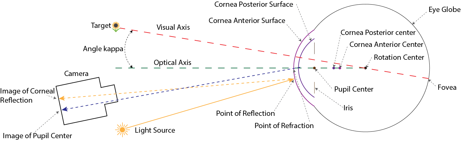
Eye Rotation
The eye’s rotational center is defined at the geometric center of the eyeball sphere. All anatomical structures are specified relative to this reference point.
Eye rotations follow Listing’s law [3], which governs the torsional component of eye movements. When the eye rotates from an initial to a final gaze direction, the rotation occurs around an axis perpendicular to both the initial and final visual axis directions.
Two rotation approaches are available:
Align the optical axis using Listing’s law, then apply foveal displacement angles (\(\alpha_{\text{fovea}}\), \(\beta_{\text{fovea}}\)) as post-rotations (default in et_simul [4]).
Compute the visual axis direction from foveal displacement and apply Listing’s law directly to this target direction.
Cornea
PyEtSimul provides both spherical and conic corneal models. In both cases, the cornea consists of two surfaces: an anterior (outer) surface and a posterior (inner) surface. The corneal center refers to the center of curvature of the anterior surface, with the posterior surface positioned relative to it based on corneal thickness.
Cornea Position
The corneal center is positioned along the optical axis, anterior to the eye’s rotation center. Its location is determined by the eyeball’s axial length and the distance from the eyeball’s front surface to the corneal center.
Spherical Cornea
The spherical model represents the anterior and posterior surfaces as concentric spheres with radii \(R_{\text{anterior}}\) and \(R_{\text{posterior}}\).
Conic Cornea
The conic model provides a more anatomically accurate representation of the human cornea [5]. The anterior and posterior corneal surfaces follow the conic formulation:
where \(R\) is the radius of curvature at the apex and \(k\) is the conic constant. Both surfaces have their own \(R\) and \(k\) parameters.
Parameter Scaling
The default anterior corneal radius is 7.98 mm. For the spherical cornea, changing the anterior radius from this default value proportionally scales related parameters—including the posterior radius, thickness offset, corneal depth, cornea position, and pupil radius—to maintain anatomical ratios. This proportional scaling does not apply to the conic cornea model.
Pupil
The pupil is positioned on the optical axis behind the corneal apex. Beyond circular pupils, PyEtSimul supports realistic non-circular shapes based on the formulation of Wyatt (1995) [6], where the pupil boundary points are generated using a Fourier-series representation:
where:
\(r_{\text{base}}\) is the base pupil radius
\(r_2\) is the elliptical component amplitude
\(\phi_{\text{major}}\) is the major-axis angle
\(r_n\) are harmonic amplitudes
\(\phi_n\) are harmonic phases
In the realistic pupil model, the pupil orientation is determined automatically based on its diameter. Wyatt (1995) observed that larger pupils in darkness (average diameter 4.93 mm) tend to have their ellipse major axes oriented vertically (0 deg), while smaller pupils in bright light (average diameter 3.09 mm) tend to have their major axes oriented horizontally (+/-90 deg). Based on this observation, 4.0 mm is set as the default threshold to classify pupils as either dilated (vertical orientation) or constricted (horizontal orientation).
Pupil Decentration
The pupil center position can shift relative to the limbus center as pupil size changes [7]. Pupil decentration is modeled using the linear decentration model of Wildenmann et al. (2013), who measured centration changes across illumination-induced pupil size variations.
The decentration offset is calculated as:
where \(d\) is the current pupil diameter, \(d_0\) is the baseline diameter, and \(\mathbf{c} = [c_x, c_y]^T\) is the coefficient vector. To simulate natural variation across a population, the coefficient vector \(\mathbf{c}\) for each simulated eye can be sampled from a normal distribution.
In addition to this built-in model, PyEtSimul features an extensible registry system that allows researchers to define and integrate their own custom decentration models.
Eyelid Occlusion
Eyelid occlusion is modeled using a geometric approach that simulates static eyelid configurations at different openness levels. Dynamic blinks are not simulated. Following observations by Nystrom et al. (2024) [8] that spontaneous blinks are dominated by upper eyelid motion while the lower eyelid remains approximately stationary, the lower edge position is fixed and only the upper eyelid position varies to control opening height.
The eyelid is modeled as a spherical surface centered at the eye’s rotation center with radius equal to half the eye axial length. The visible aperture is approximated by an ellipse in the eye’s frontal plane, with fixed horizontal width and variable vertical height. The openness parameter \(o \in [0,1]\) controls the ellipse height, where \(o=0\) represents a closed eyelid and \(o=1\) represents full opening.
A point \((x,y)\) in the eye coordinate system is visible (not occluded) if it falls inside the elliptical opening:
where \(w\) is the fixed ellipse width, \(h = o \cdot h_{\max}\) is the openness-dependent height, and \(y_{\text{center}}\) is the vertical offset that keeps the lower edge fixed.